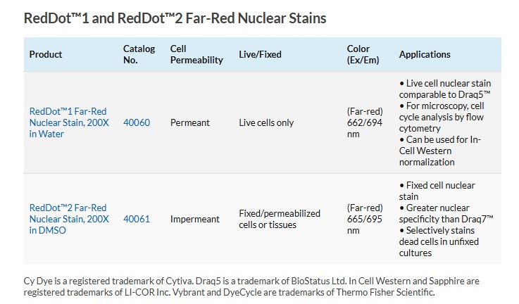
A far-red live cell nuclear stain similar to Draq5™.
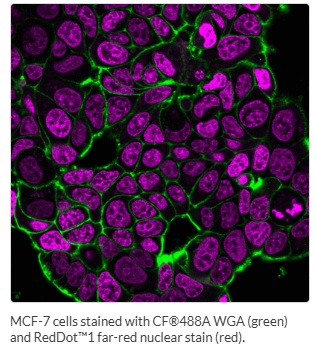
RedDot™1 is a far-red cell membrane-permeant nuclear dye similar to Draq5™. The dye is ideal for specifically staining the nuclei of live cells.
- Ideal for specifically staining the nuclei of live cells for short term imaging
- Highly thermostable and photostable, for convenient handling and demanding imaging applications
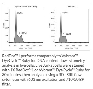
- Can be used for DNA content analysis by flow cytometry like Vybrant™ DyeCycle™ Ruby
- Can be used for normalization of In Cell Western® in fixed cells, similar to Draq5™/Sapphire®700
- λEx/λEm = 662/694 nm (with DNA), for detection in the Cy®5 channel
| Name | SKU | Size | Availability | Vendor | Price | Order | |
RedDot™1, 200X in water |
40060-T | 25 ul | Generally 1-2 weeks from receipt of order | Biotium | Log in for pricing | ||
RedDot™1, 200X in water |
40060 | 250 ul | Generally 1-2 weeks from receipt of order | Biotium | Log in for pricing | ||
RedDot™1, 200X in water |
40060-1 | 1ml | Generally 1-2 weeks from receipt of order | Biotium | Log in for pricing |
RedDot™1 can be excited at a wide range of wavelengths between 488 nm and 647 nm, including the 488 nm laser for flow cytometry, and can be used for DNA content analysis by flow cytometry. While DAPI and Hoechst dyes show a strong preference for A-T rich regions, RedDot™1 is relatively insensitive to sequence base composition.
Choose the Right Dye for Your Application
Like Draq5®, RedDot™1 is toxic to cells within a few hours, and is recommended for short term staining experiments. For a low-toxicity far-red nuclear stain for live cells, see our NucSpot™ 650 dye.
RedDot™1 can be used to stain live bacteria (gram-positive and gram-negative). Staining in live yeast is weak and not nuclear. See our Cellular Stains Table for more information on how our dyes stain various organisms.
RedDot™1 cannot be used for nuclear-specific staining in fixed cells. Please see RedDot™2 (cat# 40061), a spectrally similar dye designed for specific nuclear staining of fixed and permeabilized cells or selective nuclear staining of dead cells.
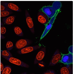
Please note: far-red dyes like RedDot™1 are not visible to the human eye, but must be imaged with a CCD camera or by confocal microscopy.
Photos

Resources
Supporting Documents:
You may also like
-
BactoSpore™ Bacterial Stains
Fluorescent dyes optimized for staining endospores. The dyes also stain both live and dead bacteria of gram-positive and gram-negative strains.
Log in for pricing -

DPX 500MG
DPX is a positively charged quencher that is often used with ANTS (90010) to study membrane fusion or permeability.
Log in for pricing -
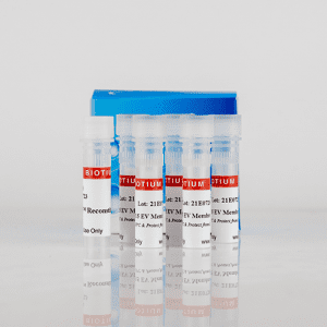
ExoBrite™ WGA EV Staining Kits
Fluorescent WGA conjugates that are optimized for bright staining of extracellular vesicles for flow cytometry.
Log in for pricing -
DiR
DiR is a lipophilic, near-infrared fluorescent cyanine dye. The dye is useful for labeling cytoplasmic membranes and has been used for near-infrared in vivo imaging.
Log in for pricing