Non-reactive mouse antibody that serves as a negative control for ExoBrite™ Flow Antibodies when detecting purified or bead-bound EVs by flow cytometry.
| Name | SKU | Size | Availability | Vendor | Price | Order | |
| ExoBrite™ IgG1 Isotype Control Flow Antibody | Generally 1-2 weeks from receipt of order | Biotium | Log in for pricing |
Product Description
ExoBrite™ IgG1 Isotype Control Flow Antibody has been found to not react with any target in human cells and have the same isotype as the ExoBrite™ Flow Antibodies against tetraspanin proteins CD9, CD63, and CD81. The antibody is available with the same conjugates as ExoBrite™ Flow Antibodies for the Pacific Blue™, FITC, PE, and APC channels.
- Non-reactive IgG1 mouse antibody
- Serves as a isotype negative control for ExoBrite™ Flow Antibodies
- Available in 5 colors
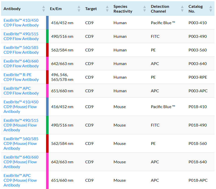
The most common proteins used as EV markers are CD9, CD63, and CD81, members of the tetraspanin family. These tetraspanins are broadly expressed on many cell types and can therefore be detected on many types of EVs, but their expression levels vary depending on the cell type of origin.
EV antibodies you can trust
Other commercially available antibodies for tetraspanin proteins CD9, CD63, and CD81 are generally not validated for isolated EVs and may require tedious optimization for your EV prep and staining protocol. The antibodies and dye labels of ExoBrite™ Flow Antibody Conjugates were carefully selected and validated for robust detection of isolated EVs. In addition, the antibodies are provided in a proprietary buffer formulation for reduced antibody aggregation and brighter EV staining for optimal accuracy and signal-to-noise. Single-color and 3-color ExoBrite™ CD9/CD63/CD81 Antibody Cocktails are also available for high-coverage EV staining or EV phenotyping, respectively.
For general EV staining, Biotium’s ExoBrite™ True EV Membrane Stains offer unparalleled coverage of EVs in a sample and address issues of dye aggregation often seen with PKH and other common membrane dyes. Biotium also offers optimized ExoBrite™ EV Surface stains conjugated to cholera toxin B (CTB), wheat germ agglutinin (WGA), and Annexin V. These stains are specially formulated for bright and specific detection of isolated EVs by flow cytometry. These ExoBrite™ EV Surface stains may also be combined with antibody staining, for multi-parameter analysis.
Biotium also offers conjugated ExoBrite™ Western Antibodies against CD9, CD63, and CD81 designed for optimal detection in EV extracts by fluorescent western blot.
Note: In our testing, we have found that ExoBrite™ 490/515 dye may bind to streptavidin coated surfaces or beads if free biotin binding sites are not blocked. We recommend performing a biotin blocking step after binding your biotinylated capture antibody to streptavidin beads or surfaces when using ExoBrite™ 490/515 conjugates. Alternatively, consider using a different ExoBrite™ dye for staining EVs captured on streptavidin beads or surfaces.
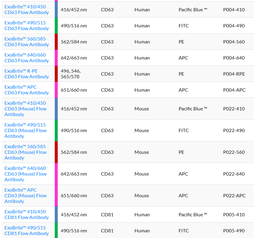
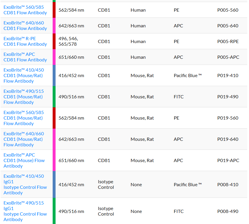
ExoBrite™ Flow Antibody Conjugates
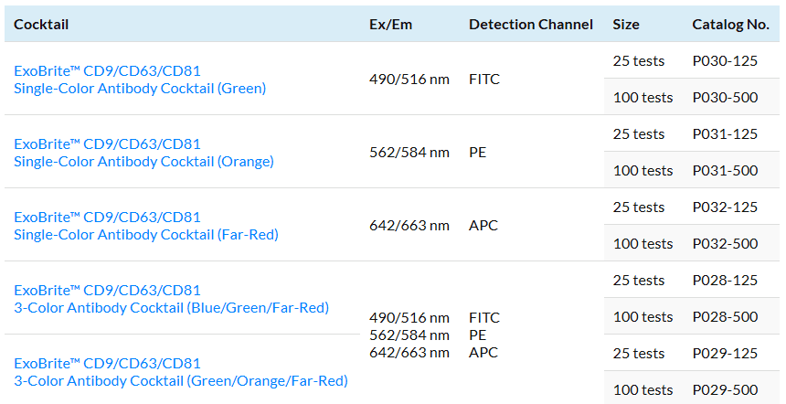
ExoBrite™ Flow Anti-Human CD9/CD63/CD81 Antibody Cocktails
Resources
Protocols
SDS
Supporting Documents
You may also like
-
Mix-n-Stain™ Nanobody Labelling Kits
These kits allow optimal labelling of single-chain nanobodies with CF® dyes or biotin in 30 minutes without a purification step.
Log in for pricing