Fluorescent cholera toxin subunit B (CTB) conjugates that are optimized for bright and clean staining of extracellular vesicles for flow cytometry.
ExoBrite™ CTB EV Staining Kits were designed to overcome some of the challenges of EV detection, particularly in flow cytometry. ExoBrite™ CTB EV Stains bind to molecules in the EV membrane for bright, specific staining, with little to no background.
Features
- Optimally formulated CTB conjugates for staining EVs
- Designed for detection by flow cytometry
- Bright signal and low background
- Stain purified or bead-bound EVs
- Compatible with antibody co-staining
- Available in 4 colors
| Name | SKU | Size | Availability | Vendor | Price | Order | |
ExoBrite™ 410/450 CTB EV Staining Kit, 500 labelings |
30111 | 500 Labellings | Generally 1-2 weeks from receipt of order | Biotium | Log in for pricing | ||
ExoBrite™ 410/450 CTB EV Staining Kit, 500 labelings |
30111-T | 100 Labellings | Generally 1-2 weeks from receipt of order | Biotium | Log in for pricing | ||
ExoBrite™ 490/515 CTB EV Staining Kit, 500 labelings |
30112 | 500 Labellings | Generally 1-2 weeks from receipt of order | Biotium | Log in for pricing | ||
ExoBrite™ 490/515 CTB EV Staining Kit, 100 labelings |
30112-T | 100 Labellings | Generally 1-2 weeks from receipt of order | Biotium | Log in for pricing | ||
ExoBrite™ 640/660 CTB EV Staining Kit, 500 labelings |
30114 | 500 Labellings | Generally 1-2 weeks from receipt of order | Biotium | Log in for pricing | ||
ExoBrite™ 560/585 CTB EV Staining Kit, 100 labelings |
30113-T | 100 Labellings | Generally 1-2 weeks from receipt of order | Biotium | Log in for pricing | ||
ExoBrite™ 560/585 CTB EV Staining Kit, 500 labelings |
30113 | 500 Labellings | Generally 1-2 weeks from receipt of order | Biotium | Log in for pricing | ||
ExoBrite™ 640/660 CTB EV Staining Kit, 100 labelings |
30114-T | 100 Labellings | Generally 1-2 weeks from receipt of order | Biotium | Log in for pricing |
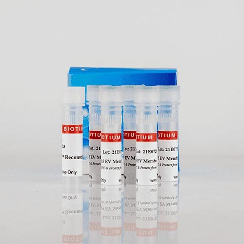
Kit Components
- ExoBrite™ CTB EV Stain
- ExoBrite™ Reconstitution Solution
- 1 vial of stain makes 100 uL of 500X staining solution
Note: The name of this product has been revised from ExoBrite™ EV Membrane Staining Kits.
ExoBrite™ CTB EV Stains are optimally formulated fluorescent conjugates of cholera toxin subunit B (CTB), which binds to GM1 gangliosides that are commonly found on the surface of mammalian lipid rafts and EVs. The stains were designed to overcome some of the challenges of EV detection, particularly in flow cytometry. Some dyes used to stain EVs can form aggregates of a similar size as exosomes or EVs, thus confounding analysis. ExoBrite™ CTB EV Stains, however, were formulated to show little to no aggregation in flow cytometry, allowing EVs to be identified with bright and specific staining. Unlike hydrophobic membrane dyes, ExoBrite™ CTB EV Stains do not bind non-specifically to polystyrene beads, meaning that they can be used to stain bead-bound EVs.
EVs are often labeled with fluorescent antibodies targeting one or more of the tetraspanin proteins CD9, CD63, and CD81. ExoBrite™ CTB staining can be combined with antibody staining, for multi-parameter analysis.
Less background and better coverage over other EV stains
ExoBrite™ CTB EV Stains were designed to offer exceptional signal:noise and more complete coverage of purified and bead-bound EVs. In the figure below, lipophilic dye DiO and plasma membrane stain CellMask™ demonstrate an unacceptable amount of dye aggregation in gated EVs. Other EV stains such as ExoFlow-ONE™ and ExoGlow™ have less coverage of EVs when compared to ExoBrite™ CTB EV Stains.
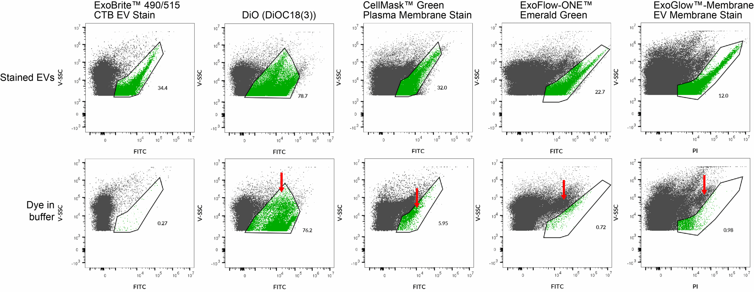
ExoBrite™ CTB EV Stains have less background and more complete staining of extracellular vesicles (EVs) than other classic and competitor dyes. EVs were purified from MCF-7 cell supernatant using size exclusion chromatography (SEC). The purified EVs were stained in PBS with the indicated dyes (top row). The stained EV population was gated, with the number showing the percentage of particles falling within the exosome gate. Each dye was also added to filtered PBS (bottom row), to look for dye aggregation and non-specific background. Red arrows indicate these dye aggregates. Lipophilic dyes like DiO show a high number of particles of similar size to EVs, making them unsuited for small particle staining. CellMask™ also shows an unacceptable amount of dye aggregation falling within the EV gate. The ExoFlow-ONE™ and ExoGlow™ dyes form aggregates that can mostly be gated away from the EVs, but they also show less-complete coverage of EVs than ExoBrite™ CTB stains. (Click to enlarge).
Notes:
- ExoBrite™ CTB EV Stains have been found to label EVs derived from several tested cell lines (see Validated EV Sources below), but may not stain EVs from every source.
- In our testing, we have found that ExoBrite™ 490/515 dye may bind to streptavidin coated surfaces or beads if free biotin binding sites are not blocked. We recommend performing a biotin blocking step after binding your biotinylated capture antibody to streptavidin beads or surfaces when using ExoBrite™ 490/515 conjugates. Alternatively, consider using a different ExoBrite™ dye for staining EVs captured on streptavidin beads or surfaces.
Resources
Protocols
SDS
Supporting Documents
You may also like
-
Extracta DNA Prep for PCR
Extracta DNA Prep for PCR is a two-component reagent kit for rapid extraction of PCR-ready genomic DNA from a variety of tissues. Samples are processed in less than 30 minutes with minimal hands-on time and technical skill.
Log in for pricing -
QuickExtract™ DNA Extraction Solution
QuickExtract™ DNA Extraction Solution is a fast and simple method for extracting PCR-ready genomic DNA from almost any sample for screening and genotyping applications.
Log in for pricing -
QuickExtract™ Plant DNA Extraction Solution
Rapid and efficient extraction of PCR-ready genomic DNA from plant leaf samples
Log in for pricing -
Red Cell Lysis Solution
Selectively lyse red blood cells while leaving white blood cells intact. To be used with the MasterPure Kits.
Log in for pricing